DetectionEntry 03.3
Ultrasound and MRI
What was established
Two detection methods that work without ionising radiation and see things mammography cannot — because physics, not equipment, is the real difference.
Sound and magnetism, solving different problems
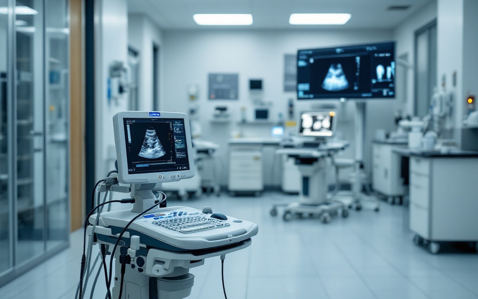
Ultrasound sends high-frequency sound waves into tissue and reads the returning echoes. Fluid-filled structures, such as a simple cyst, reflect the waves in a distinct pattern; solid masses behave differently. That physical distinction is what made ultrasound useful almost immediately after X-ray mammography became standard: when a mammogram flagged something that might be a cyst or a solid lump, ultrasound could characterise it without additional radiation. The technology requires no compression and no ionising dose, which is why it spread quickly into breast clinics from the late 1970s onward.
Its limits are the same as its strengths. Ultrasound is good at interrogating a known target but poorly suited to surveying large volumes of tissue systematically. Dense breast tissue does not obstruct sound the way it blocks X-rays, yet the images are operator-dependent and covering the entire breast reproducibly remains technically demanding. Automated whole-breast ultrasound addresses some of this, though the evidence base for population use is still being assembled.
Lifted out of the flow
Detection methods at a glance
- Ultrasoundreads reflected sound echoes; no radiation; best for characterising a known target or distinguishing cyst from solid mass
- MRIreads magnetic resonance signal from hydrogen nuclei; uses gadolinium contrast to reveal blood-vessel proliferation; no ionising radiation
- Mammographyreads differential X-ray absorption; gold standard for population screening but limited in dense tissue
Magnetic resonance imaging works on an entirely different principle. A strong magnetic field aligns the hydrogen nuclei in water molecules throughout the body; radiofrequency pulses tip them out of alignment; the signal emitted as they return is recorded and reconstructed into images. Breast MRI is almost always performed with an intravenous gadolinium-based contrast agent, which accumulates in areas where blood vessels are proliferating — a characteristic of many malignant tumours. That enhancement pattern, rather than any structural shadow, is what MRI is reading.
The consequence is that MRI is highly sensitive — it finds things — but specificity is more variable, meaning it also generates findings that prove benign on biopsy. Its established role, confirmed by a series of observational studies and reviewed periodically by bodies including the International Agency for Research on Cancer, is surveillance of women carrying high-risk gene variants, where the greater sensitivity justifies the higher false-positive rate. It is also used to assess tumour extent before surgery and to evaluate the response of a tumour to chemotherapy given before the operation.
Neither method replaces the other, and neither replaces mammography for population screening. What each offers is a different physical window into the same tissue — echoes where mammography casts shadows, magnetic resonance where density is irrelevant — and clinical practice uses all three according to what question needs answering.
Elsewhere in detection
- Mammographic techniqueRobert Egan standardised exposure and positioning in the late 1950s, which is what turned an unreliable picture into a usable one.
- Film to digitalReplacing film with a detector changed the dose, the storage and what could be done to the image afterwards.
- The pathology slideThe final answer is a piece of tissue on glass, stained, and looked at by a person.