DetectionEntry 03.1
Mammographic technique
What was established
How a standardised protocol in the late 1950s converted an interesting but unreliable picture into a diagnostic tool that could be taught, repeated, and trusted.
The problem with the early pictures
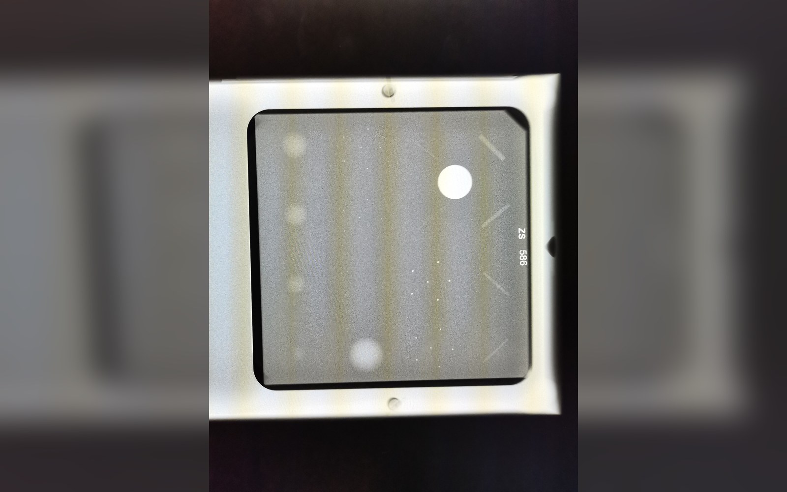
X-rays had been turned on breast tissue almost as soon as Wilhelm Röntgen announced his discovery in 1895, and the pictures that followed across the first half of the twentieth century were adequate proof that something was happening in there. Surgeons in Germany and Uruguay made early attempts in the 1910s and 1920s, and by the 1930s a handful of radiologists were producing images that could, under the right conditions, reveal gross masses. The phrase "under the right conditions" is where the whole endeavour stalled. Technique varied wildly between operators and institutions. Exposure times, kilovoltage, the type of film, the degree of compression applied, the positioning of the patient — all of it was improvised, and the results were correspondingly inconsistent. A radiologist who had produced a convincing image of a tumour one week could not reliably reproduce it the next, and the images produced at one hospital bore little systematic relationship to those produced at another. The breast, being largely soft tissue with relatively low inherent contrast, demanded much finer calibration than the chest or limb radiography that had become routine. Nobody had yet worked out what that calibration was.
The word mammography itself was already in circulation, but it described a category of attempt rather than a defined procedure. What was missing was not the physics — the physics was well understood — but a reproducible protocol: a specified combination of equipment settings, film type, processing chemistry, compression force and patient positioning that would yield, consistently and in different hands, an image of sufficient quality to support a diagnostic judgement. That is what Robert Egan supplied.
Lifted out of the flow
Key steps in the standardisation process
- Pre-Egan mammographyvariable kilovoltage, film types and positioning; no agreed protocol; results inconsistent between operators and institutions
- Egan's 1960 protocollow-kVp exposure (~26–28 kVp), industrial-grade fine-grain film, specified processing, controlled compression and positioning
- Multi-centre validationprogramme supported by the National Cancer Institute; 24 institutions tested the protocol; reproducibility confirmed
- Dedicated units (1960s–70s)purpose-built mammography equipment replaced adapted general radiography machines; geometry and focal spot refined
- Screen-film era (from early 1970s)dose reduced substantially while resolution maintained; American College of Radiology begins accreditation work
- Mammography Quality Standards Act (1992)US legislation mandating facility accreditation and minimum technical performance criteria
What Egan established
Egan was a radiologist working at the M. D. Anderson Hospital in Houston, Texas, in the late 1950s — an institution already developing a serious interest in systematic cancer investigation. His approach was methodical in a way that previous work had not been. Rather than accepting that breast radiography was inherently difficult and its results inherently variable, he treated it as an engineering problem: identify the parameters that produce a useful image, write them down precisely, and then verify that others can reproduce the result when following the same written protocol.
The protocol Egan developed and published in 1960 specified low-kilovoltage exposure — around 26 to 28 kVp — which maximises the photoelectric effect and thus the contrast between tissues of similar density. He used industrial-grade film rather than the standard medical film then in common use, which gave finer grain and better resolution of the microcalcifications and soft-tissue masses that are the radiological signatures of early disease. He specified processing conditions, positioning, and a degree of compression that flattened the breast sufficiently to bring structures at different depths into the same focal plane without distorting them beyond recognition. The whole package was documented, testable, and transferable.

The validation was not just self-reported. The National Cancer Institute supported a programme in which Egan's protocol was sent to other centres and assessed against their results. Twenty-four institutions tested it, and the output was consistent enough to establish that the method could travel — that it was a protocol rather than a personal skill. That is the moment mammography became a reproducible technique rather than an intermittent achievement.
The clinical results Egan reported were striking for the time. In a series published through the early 1960s, he demonstrated that the method could identify lesions — including clusters of microcalcifications — that were too small to be palpated and that had not been clinically suspected. The critical finding was not merely that the images were clearer; it was that they carried information unavailable by any other means then in routine use. A substantial proportion of the cancers he identified in his early series were what would later be classified as carcinoma in situ or small invasive tumours, precisely the cases where earlier detection might alter the eventual course of disease.
The critical finding was not merely that the images were clearer; it was that they carried information unavailable by any other means then in routine use.
Standardisation and its consequences
The effect of having a written, validated protocol cascaded outward in ways that went beyond improved imaging. Once technique was standardised, it became possible to train radiologists and radiographers in a systematic way rather than through apprenticeship to a particular practitioner. It became possible to compare images taken at different times in the same patient, because the parameters were fixed. And it became possible, eventually, to design a population trial in which one group received regular mammographic examination and another did not — and to trust that the imaging being delivered in the trial arm was consistent enough to constitute a real intervention.
That last consequence mattered enormously. The randomised trials of mammographic screening that began in the 1960s with the Health Insurance Plan study in New York, and continued through the Swedish trials of the 1970s and 1980s, all depended on the assumption that the mammography being delivered was reliable and reproducible. Without a defined technique to specify in a protocol, a trial would be testing not screening but screening-as-performed-variably-by-whoever-happened-to-be-available, which is not the same thing at all. Egan's work is the precondition for the evidence base.
Equipment continued to evolve after Egan's foundational publications. Dedicated mammography units replaced general radiographic equipment through the 1960s and 1970s, allowing further refinement of geometry and focal spot size. Screen-film combinations developed from the early 1970s onward dramatically reduced radiation dose while maintaining resolution, a development coordinated in part through the work of the American College of Radiology, whose accreditation standards eventually codified what Egan had pioneered informally. The Mammography Quality Standards Act, passed in the United States in 1992, gave those standards legal force, requiring accreditation of all facilities offering mammographic screening and specifying minimum technical performance criteria in a way that traces a direct line back to the logic of Egan's original project.
Compression, consistently one of the less comfortable aspects of the examination for the patient, remained technically non-negotiable throughout these decades. It is not punitive engineering but a physical requirement: reducing tissue thickness equalises exposure across the image, reduces the radiation dose needed to penetrate the breast, minimises geometric blur from patient movement, and separates overlapping structures that would otherwise obscure one another. The final technical parameters of modern analogue and then digital systems differ substantially from Egan's 1960 specification, but the underlying reasoning — that image quality is a function of controlled, documented, reproducible conditions rather than operator intuition — is unchanged.
Lifted out of the flow
Chronology
- 1895Röntgen announces X-ray discovery; early breast imaging attempts follow within years
- 1910s–1930sisolated breast X-ray work in Germany and Uruguay; no standardised approach
- Late 1950sRobert Egan develops his protocol at M. D. Anderson Hospital, Houston
- 1960Egan publishes the standardised technique; multi-centre validation follows
- 1963Health Insurance Plan trial, New York, begins: first randomised screening trial, dependent on reproducible technique
- 1992Mammography Quality Standards Act passed in the United States
The transition from film to digital detectors from the late 1990s onward changed the storage, transmission and post-processing possibilities of the image without disturbing the foundational logic of standardised acquisition.
Elsewhere in detection
- Film to digitalReplacing film with a detector changed the dose, the storage and what could be done to the image afterwards.
- Ultrasound and MRIOne reads echoes and the other listens to atoms in a magnetic field; neither casts a shadow, which is why they see different things.
- The pathology slideThe final answer is a piece of tissue on glass, stained, and looked at by a person.