TreatmentEntry 06.1
Radiotherapy
What was established
After the surgeon finishes, directed radiation reduces the chance that residual cells will regrow — and deciding exactly where to aim it is a geometry problem as much as a biological one.
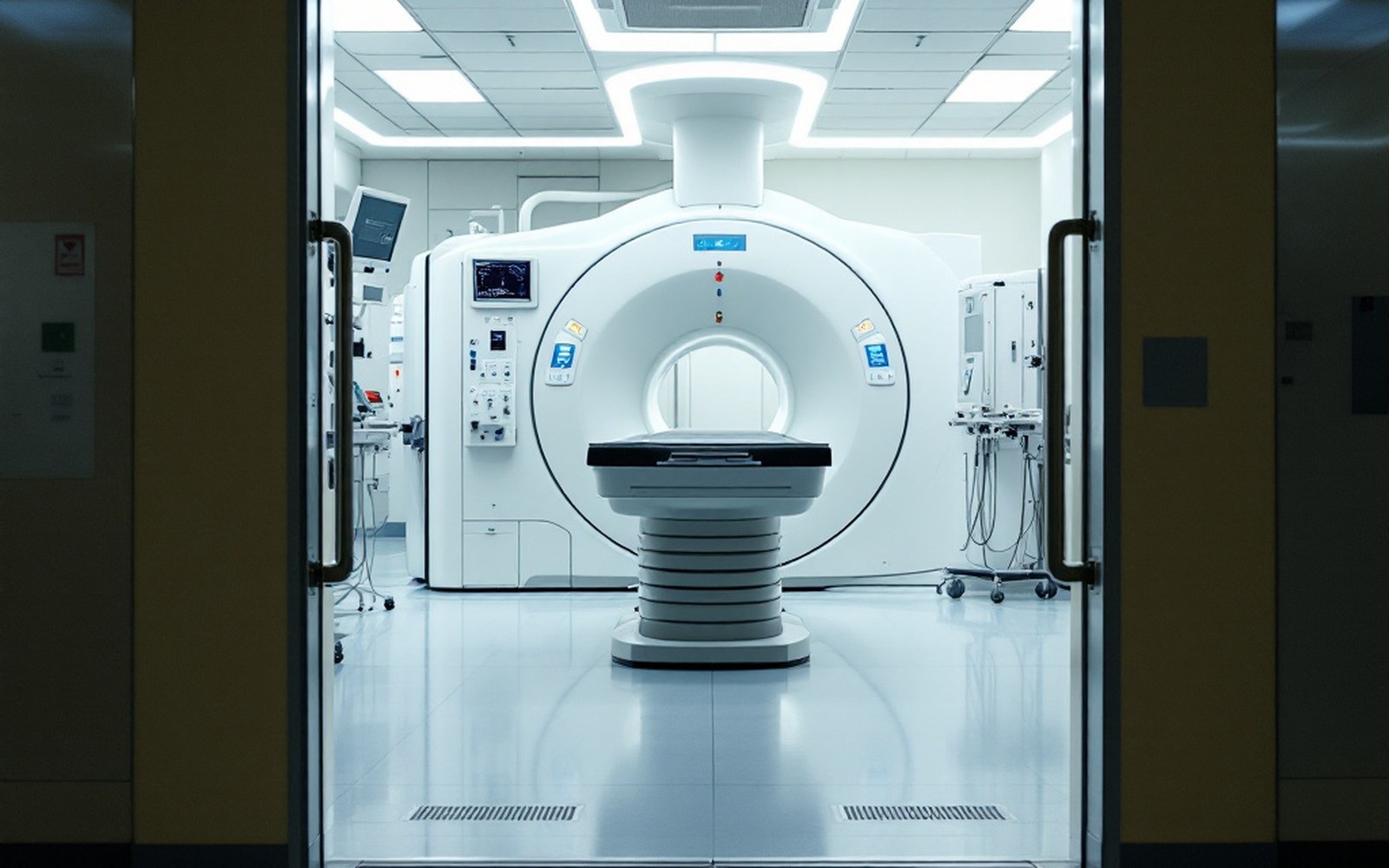
From empirical treatment to precise field design
Radiation was applied to breast tumours within a few years of Wilhelm Röntgen's discovery of X-rays in 1895, long before anyone understood the mechanism. The effects were visible but inconsistent, and for the first half of the twentieth century radiotherapy sat in an uneasy relationship with surgery — sometimes given before an operation to shrink a mass, sometimes after, sometimes instead. Geoffrey Keynes, working in London in the 1920s and 1930s, used interstitial radium needles alongside conservative excision in a deliberate challenge to the radical mastectomy then dominant; his results were respectable but his surgical colleagues were largely unconvinced, and the evidence base for what he was doing rested on observation rather than randomisation.
What changed the situation was the randomised trial. The NSABP B-06 trial, run by the National Surgical Adjuvant Breast and Bowel Project from Pittsburgh, Pennsylvania, showed that lumpectomy with radiotherapy produced survival equivalent to mastectomy in women with tumours of four centimetres or less. The radiotherapy arm was essential to that equivalence: lumpectomy alone, also included in the trial, produced substantially higher local recurrence rates. The Milan I trial reached the same conclusion independently. Together they established adjuvant radiotherapy — radiation added after surgery — as the standard accompaniment to conserving surgery.
Lifted out of the flow
Chronology
- 1895Röntgen discovers X-rays; radiation applied to tumours within years
- 1920s–30sGeoffrey Keynes uses radium needles with conservative excision in London
- NSABP B-06 and Milan I (reported 1980s)establish lumpectomy plus radiotherapy as mastectomy-equivalent
- 2008UK START trials report, hypofractionation shown equivalent to conventional schedule
- ~2020FAST-Forward trial, five-fraction course in one week, non-inferior outcomes
What the field covers and how it is calculated
The geometry matters because the breast is not a uniform target. After lumpectomy, the principal concern is the tumour bed — the cavity left by removal — and the surrounding tissue where residual microscopic disease is most likely. Standard whole-breast irradiation treats the entire remaining breast tissue to a base dose, typically followed by a boost to the tumour bed delivering an additional increment. After mastectomy, radiotherapy is directed at the chest wall and sometimes the regional lymph node basins, depending on nodal involvement and tumour characteristics at the time of surgery.
Planning is done with CT imaging. The radiation oncologist delineates target volumes — the tissue that should receive full dose — and organs at risk, principally the heart and the ipsilateral lung, which sit immediately behind the breast and receive incidental exposure on every field. Reducing cardiac dose has been a sustained engineering and dosimetric project for decades, driven by evidence from the Early Breast Cancer Trialists' Collaborative Group that radiotherapy's gains in local control are real but must be weighed against late cardiac toxicity, particularly for left-sided tumours. Breath-hold techniques — the patient inhales and holds while the beam is active, displacing the heart posteriorly — are one approach now widely used to manage this.

Dose and fractionation have both been revised substantially by trial evidence. The UK START trials, reported from 2008 onward, demonstrated that hypofractionation — delivering a higher dose per fraction over fewer sessions — gave local control and toxicity outcomes equivalent to the conventional long course, and it subsequently became the standard schedule in the United Kingdom and then in many other systems. A further compression, ultra-hypofractionation or FAST-Forward, was shown in trials published around 2020 to deliver five fractions over a single week with non-inferior outcomes, reducing treatment burden substantially.
Partial-breast irradiation, which restricts the treated volume to the immediate tumour bed region, has also been tested in trials as a strategy for selected patients with early, low-risk disease, on the biological argument that recurrence predominantly occurs near the original site. Results have been mixed enough that it remains a clinical research question rather than a settled standard in most systems.
After lumpectomy, the principal concern is the tumour bed — the cavity left by removal — and the surrounding tissue where residual microscopic disease is most likely.
What radiation does — killing or sterilising cells through ionising damage to DNA — has been understood for decades. What took time was establishing exactly how much, to where, over how long, and in whom it helps without doing disproportionate harm elsewhere.
Lifted out of the flow
Concepts in this article
- Adjuvant radiotherapyradiation given after surgery to reduce local recurrence, not as primary treatment
- Tumour bed boostextra dose to the cavity left by lumpectomy, in addition to the whole-breast dose
- Hypofractionationfewer fractions at higher dose per session; equivalent outcomes, shorter course
- Organs at riskheart and ipsilateral lung, delineated in planning to limit incidental exposure
- Breath-hold techniquepatient holds inhaled breath during beam delivery to displace heart from the field
Elsewhere in treatment
- Cytotoxic chemotherapyCombination regimens came out of trial programmes rather than from any single laboratory insight.
- TamoxifenA compound developed for something else entirely became the long-running hormonal treatment, and the receptor work is what explained why it worked.
- Aromatase inhibitorsBlocking the enzyme rather than the receptor is a different mechanism with different consequences.