The diseaseEntry 01.1
Ducts and lobules
What was established
The breast is not a uniform mass — it is an organised, branching architecture, and the kind of growth that forms, and what it does next, is inseparable from where in that architecture it begins.
The tree and its terminals
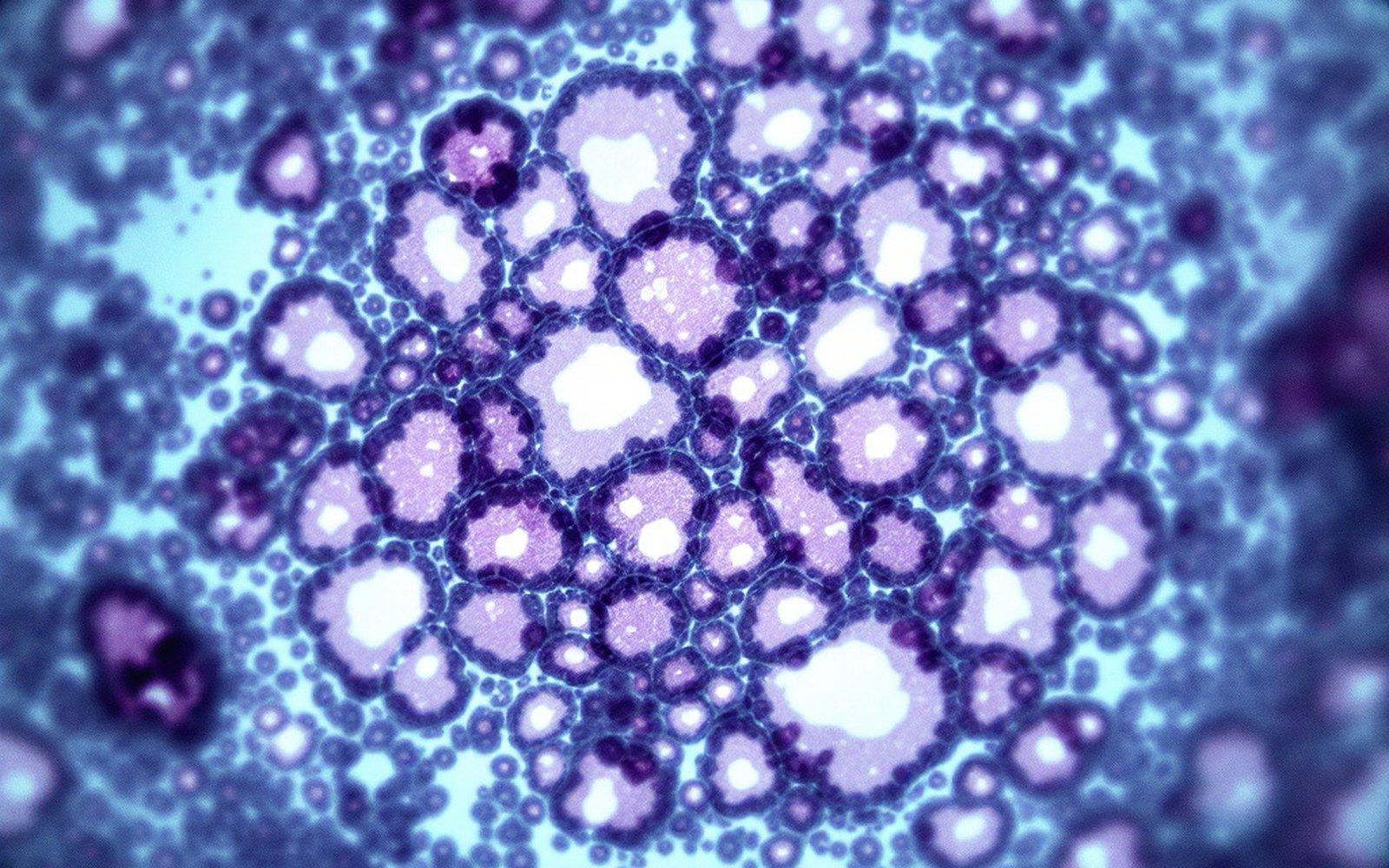
The functional unit of the breast is a system that looks, in textbook diagrams, like a tree drawn upside down. At the apex sits the nipple. From it, between fifteen and twenty lactiferous ducts fan outward and inward through the fatty and fibrous stroma that gives the organ its bulk. Each major duct branches progressively into smaller ducts, and those smaller ducts terminate in clusters of tiny secretory sacs called lobules. The lobule and its draining duct together form the terminal duct lobular unit, a phrase that appears throughout pathology literature because it is the site where the overwhelming majority of breast tumours originate. The nipple and the larger collecting ducts exist mainly as conduits; the action, both normal and pathological, happens at the far end of the tree.
The lobules themselves change through life. They are classified, by convention, into three types — Type 1 lobules are the sparse, undeveloped structures present before puberty and in postmenopausal women; Type 2 and Type 3 are progressively more elaborated, proliferating under the influence of oestrogen and progesterone across the reproductive years and becoming maximally developed during pregnancy and lactation, when the epithelial cells that line them are producing milk. After weaning, the lobules involute back toward simpler forms. This cycling matters because cells that are actively dividing accumulate copying errors, and the tissues most subject to hormonal stimulation across a lifetime carry the most accumulated opportunity for mutation.
Lifted out of the flow
Structural glossary
- Terminal duct lobular unitthe junction of a small duct and its cluster of lobules; the commonest site of tumour origin
- Luminal cellsthe inner epithelial layer of the duct or lobule, facing the hollow centre
- Myoepithelial cellsthe outer epithelial layer; sits on the basement membrane and contracts during lactation
- Basement membranethe structural boundary separating epithelium from stroma; its integrity defines "in situ" vs invasive
- E-cadherinadhesion protein characteristically lost in lobular carcinoma; its absence shapes the single-file growth pattern
- Lobule types (1–3)a developmental classification reflecting the degree of lobular elaboration across the lifespan
The epithelium lining the ducts and lobules is two-layered. An inner layer of luminal cells — the cells that face the hollow centre of the duct or lobule — is surrounded by an outer layer of myoepithelial cells. The myoepithelial layer sits on a basement membrane, which is the structural boundary that separates the epithelium from the surrounding stroma. This boundary is the line that defines whether a lesion is confined or has become invasive carcinoma. When cells proliferate abnormally but stay within it, the result is carcinoma in situ, a classification with significant consequences for everything that follows. When they breach it, the biology, the behaviour and the clinical situation all change.
Ductal and lobular: what the names mean
Because the terminal duct lobular unit sits at the junction of two named structures, tumours arising there have historically been divided into ductal and lobular types — a classification that remains in clinical use even though the original anatomical logic behind it has been substantially revised. The distinction was formalised through twentieth-century pathology, and it tracks real biological differences. Invasive lobular carcinoma, which accounts for roughly ten to fifteen percent of invasive breast cancers according to data maintained by the International Agency for Research on Cancer, has a characteristic growth pattern: its cells infiltrate the stroma in single files rather than forming the compact masses typical of ductal tumours. This makes it harder to see on mammography, harder to feel, and in some studies harder to detect until it has reached a larger size.
The cellular basis for this infiltrative pattern lies in a molecule called E-cadherin, a protein that normally keeps epithelial cells adherent to one another. Invasive lobular carcinoma characteristically loses E-cadherin expression — a finding now incorporated into pathological diagnosis — and this loss of adhesion is what allows the cells to disperse through tissue in columns rather than clumps. Ductal tumours retain E-cadherin and behave differently at a structural level, even before receptor status, grade or stage is considered. Those latter features are layered on top of the ductal-or-lobular distinction rather than replacing it.

Within the ductal category there is further heterogeneity. Ductal carcinoma in situ — DCIS — presents as abnormal cells filling and expanding ducts, and it is classified by its architectural pattern (solid, cribriform, micropapillary) and by nuclear grade. These subtypes carry different likelihoods of progression to invasive disease, which is where the clinical and epidemiological complexity of DCIS becomes most acute. The question of how much DCIS would ever become invasive if left alone is central to ongoing debates about overdiagnosis in populations offered screening, a debate in which the Cochrane Collaboration and the US Preventive Services Task Force have both contributed careful, and sometimes divergent, analyses.
Why the architecture matters downstream
The architecture of ducts and lobules is not merely anatomical classification for its own sake. It is the substrate from which every subsequent layer of understanding grows. Receptor biology, for instance, operates at the level of the epithelial cell: the oestrogen receptor is expressed in the luminal cells of the duct and lobule, and its presence or absence in a tumour determines whether hormonal treatments are relevant. HER2 overexpression similarly occurs in the luminal epithelium. A tumour classified by receptor status is, at root, a tumour of cells that came from a specific layer in a specific part of the tree.
Surgical reasoning was also shaped by this anatomy for most of the twentieth century, though the relationship between anatomical reasoning and surgical radicalism was complicated. William Halsted conceived his radical mastectomy in part as a response to what he believed about the local spread of disease through tissue planes, but the architecture of the ductal system also informed debates about whether removing more breast tissue offered any benefit. When Bernard Fisher and the National Surgical Adjuvant Breast and Bowel Project ran the trials that challenged radical surgery in the 1970s and 1980s, the underlying question was partly about whether the ductal tree needed to be excised in its entirety, or whether the biology of spread made that premise wrong.
The cellular basis for this infiltrative pattern lies in a molecule called E-cadherin, a protein that normally keeps epithelial cells adherent to one another.
The lobular architecture also explains one specific feature of the disease that has clinical consequences: lobular carcinoma's tendency to spread to unusual metastatic sites — the gastrointestinal tract, the peritoneum, the meninges — compared with ductal tumours, which more often seed the liver, lung and bone. This tropism is not fully understood mechanistically, but it is consistent enough to be noted in WHO classification criteria for tumours of the breast and to influence the monitoring of patients with lobular histology.
The ducts and lobules, then, are the origin point — anatomically, biologically and historically — for almost the entire body of knowledge that accumulated around this disease in the twentieth century. The tissue is the subject, and understanding its architecture is the precondition for understanding what happens when it goes wrong.

Lifted out of the flow
Key distinction to call out
- Ductal vs lobular: not simply where the cell came from, but a distinction that predicts growth pattern (mass vs single file), mammographic visibility, and metastatic behaviour
Elsewhere in the disease
- Carcinoma in situCells confined within the duct wall are classified separately from those that have crossed it, and that boundary is the whole basis of staging.
- Invasive carcinomaOnce the basement membrane is breached the classification changes and so does everything downstream of it.
- Grade and stageGrade describes how the cells look and stage describes how far they have gone; the two are routinely confused and mean different things.