DetectionEntry 03.2
Film to Digital
What was established
Replacing film with a detector changed dose, storage, and what could be done to the image afterwards.
The physics of the swap
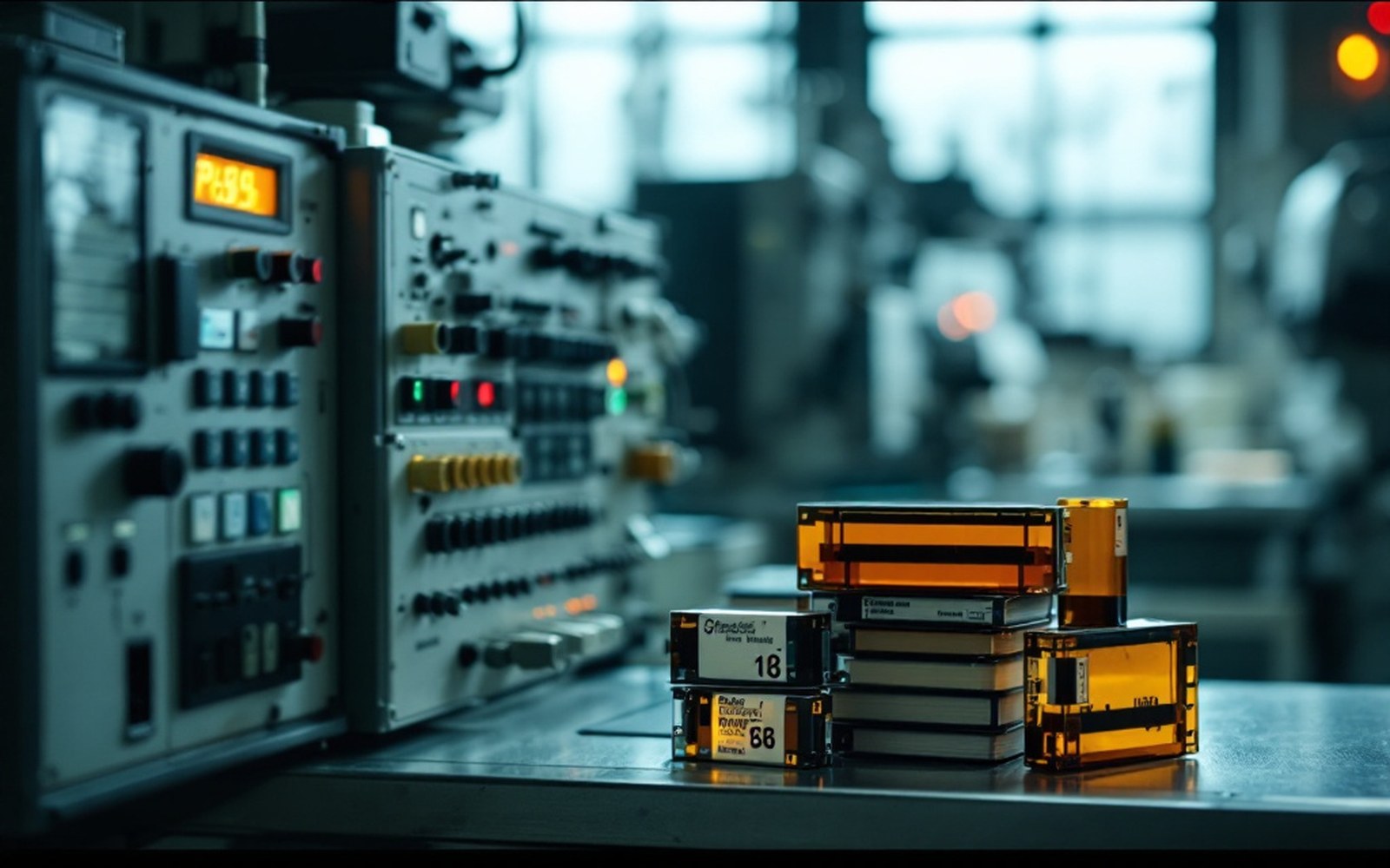
Mammographic film works by the same principle that governed diagnostic radiology for most of the twentieth century: X-ray photons strike a silver-halide emulsion, trigger a chemical reaction, and leave a latent image that developer solution renders visible. The system is cheap and produces a fine-grained picture, but it is a dead end. Once fixed, the image cannot be brightened, windowed, or transmitted without physically moving the film.
Digital detectors broke that constraint. The two main technologies that reached clinical use are computed radiography (CR), which uses a photostimulable phosphor plate scanned by a laser after exposure, and full-field digital mammography (FFDM), which uses a flat-panel detector — typically amorphous selenium — that converts X-ray photons directly into an electrical signal. FFDM captures data in a single step; CR is an intermediate technology that fits into existing film-based workflows before full replacement. Both produce a numerical matrix of pixel values rather than a fixed image, which is the essential difference.
Lifted out of the flow
Key technologies
- Film-screen mammographysilver-halide emulsion; fixed image; dominant until the 2000s
- Computed radiography (CR)phosphor plate scanned by laser; transitional technology
- Full-field digital mammography (FFDM)flat-panel detector; single-step digital capture
- PACSPicture Archiving and Communication System; digital image storage and retrieval
- DICOMfile format standard enabling cross-system image exchange
- Tomosynthesisarc-sweep X-ray plus reconstruction algorithm; produces slice images; requires digital chain
What changed in practice
The numerical image can be post-processed: contrast adjusted, edges enhanced, areas magnified — all after the patient has left the room. Radiologists can view the image on a calibrated monitor at whatever window level suits the tissue in front of them, rather than holding a light-sheet under fixed illumination. That flexibility matters because dense breast tissue and fatty tissue require different display conditions to reveal subtle findings.
Radiation dose proved more nuanced than early optimism suggested. Digital detectors are generally more efficient at capturing X-ray photons than film-screen combinations, which in principle allows a lower dose for equivalent image quality. The Digital Mammographic Imaging Screening Trial (DMIST), a large American study published in 2005 and run across multiple institutions in the United States and Canada, found that FFDM performed similarly to film-screen mammography overall, but significantly better in women under fifty, those with dense breasts, and pre- or perimenopausal women — the groups in whom film had always performed least well. Mean glandular dose varied across sites and systems; the trial did not establish digital as uniformly lower-dose, but subsequent equipment optimisation narrowed the gap further.
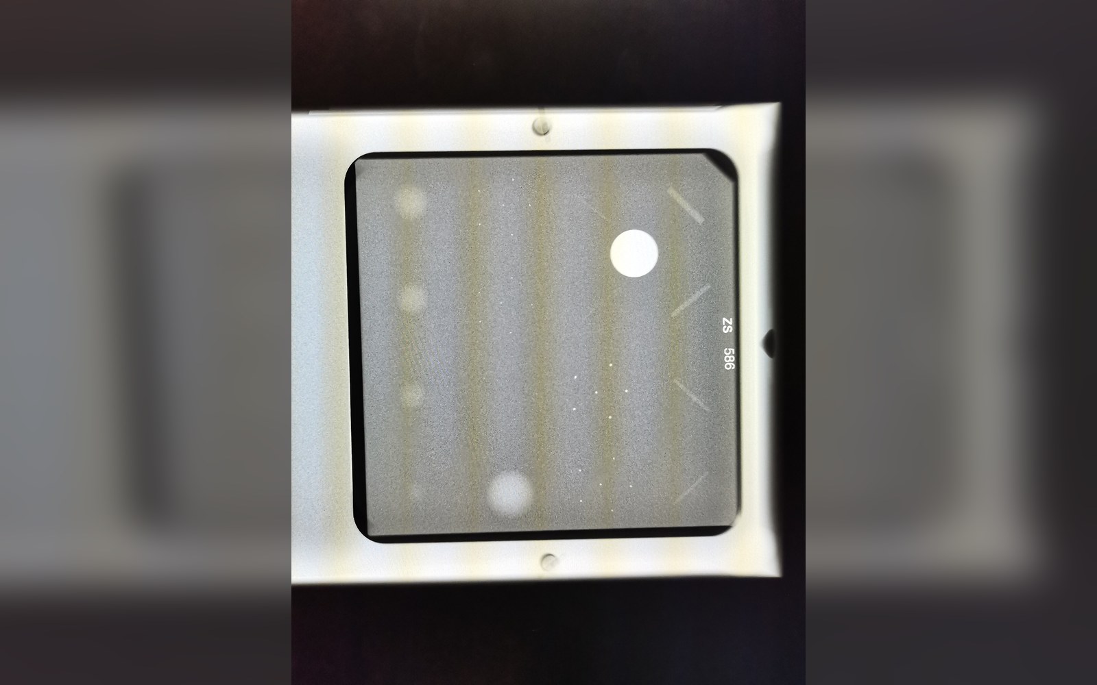
Storage changed radically. A film archive requires physical space, environmental controls, and retrieval staff. Digital images enter a Picture Archiving and Communication System (PACS), where they can be retrieved instantly, duplicated without loss, and transmitted to a remote reader over a network. The standardised file format, DICOM (Digital Imaging and Communications in Medicine), allows images taken on one manufacturer's equipment to be read on another's. That interoperability enabled teleradiology — off-site reading of images — and made centralised double-reading in national screening programmes more practical.
The most consequential downstream development from digitisation is tomosynthesis: a system in which the X-ray tube sweeps an arc around the breast and a reconstruction algorithm assembles the acquired projections into a stack of thin slices, effectively removing the superimposition of overlapping tissue that masks lesions on conventional two-dimensional mammography. Tomosynthesis is not possible with film; it depends entirely on the digital signal chain. Several large studies, including the Oslo Tomosynthesis Screening Trial published in 2013, reported higher cancer detection rates and lower recall rates compared with two-dimensional digital mammography. Whether the additional cancers detected represent genuine benefit or contribute to overdiagnosis — the detection of cancers that would not have harmed the patient — remains an active question, one the Cochrane Collaboration and other reviewers have not yet resolved.
The numerical image can be post-processed: contrast adjusted, edges enhanced, areas magnified — all after the patient has left the room.
The transition from film to digital was, in one sense, a change of medium. In another, it was the opening of an entirely different technical era: images that could be computed, not just looked at.
Lifted out of the flow
Chronology
- 2005DMIST results published; FFDM shown superior in dense-breast and younger populations
- 2013Oslo Tomosynthesis Screening Trial reports higher detection and lower recall rates
- Ongoingwhether tomosynthesis increases overdiagnosis remains unresolved
Elsewhere in detection
- Mammographic techniqueRobert Egan standardised exposure and positioning in the late 1950s, which is what turned an unreliable picture into a usable one.
- Ultrasound and MRIOne reads echoes and the other listens to atoms in a magnetic field; neither casts a shadow, which is why they see different things.
- The pathology slideThe final answer is a piece of tissue on glass, stained, and looked at by a person.