DetectionEntry 03.4
The Pathology Slide
What was established
A biopsy moves tissue from body to laboratory, and the diagnosis lives in what a trained eye reads from a stained section on glass.
What the slide actually shows
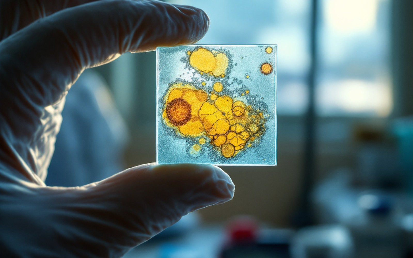
Tissue removed by needle or scalpel reaches the pathology laboratory fixed in formalin, embedded in paraffin wax, sliced by a microtome to a thickness of a few micrometres, and mounted on a glass slide. The standard stain is haematoxylin and eosin — haematoxylin colours nuclei blue-purple, eosin colours cytoplasm pink — a combination in routine use since the late nineteenth century. Under the microscope, a pathologist reads architecture and cellular detail simultaneously: whether ducts are intact or breached, how abnormal the nuclei look, how often cells are dividing.
These observations translate directly into clinical categories. Grade and stage begin here, on the slide — grade assessed by nuclear pleomorphism, tubule formation and mitotic count, using the Elston-Ellis modification of the Bloom-Richardson system formalised in the 1990s. A cell confined within the duct wall is carcinoma in situ; one that has crossed the basement membrane is invasive, a distinction with real consequences for everything that follows. The pathologist also notes tumour size, margin status, and whether lymphatic channels contain tumour cells — each item feeding into the staging calculation.
Lifted out of the flow
Key steps: biopsy to diagnosis
- Formalin fixationhalts cellular decay immediately after removal
- Paraffin embeddingsupports tissue for thin sectioning
- Microtomycuts sections typically 3–5 µm thick
- H&E staininghaematoxylin (nuclei) + eosin (cytoplasm); the baseline view
- Immunohistochemistryantibody-based stains for ER, PR, HER2 and other markers
- Pathologist reviewthe interpretive step that produces the formal diagnosis
Haematoxylin and eosin alone is not always enough. Immunohistochemistry applies antibodies directly to the section: antibodies against the oestrogen receptor mark the nuclei of hormone-sensitive tumours brown, and the result — positive or negative, and scored by the percentage of stained cells — determines whether endocrine therapy is likely to work. HER2 status is assessed the same way, with reflex testing by in-situ hybridisation when the initial stain is equivocal. The World Health Organization's classification of tumours, revised most recently in 2019, standardises these categories internationally.
None of this is automated in any final sense. Image-analysis software assists with mitotic counts and receptor scoring, but the interpretive act — recognising an unusual growth pattern, deciding a margin is close, reconciling a complex immunoprofile — remains with the pathologist. The slide is where measurement meets judgement, and the treatment plan follows from what that judgement concludes.
Elsewhere in detection
- Mammographic techniqueRobert Egan standardised exposure and positioning in the late 1950s, which is what turned an unreliable picture into a usable one.
- Film to digitalReplacing film with a detector changed the dose, the storage and what could be done to the image afterwards.
- Ultrasound and MRIOne reads echoes and the other listens to atoms in a magnetic field; neither casts a shadow, which is why they see different things.